


Fluorescence microscopy is a cornerstone of modern life sciences, enabling researchers to track molecular interactions, study live-cell dynamics, and decode disease mechanisms. However, achieving high-quality images requires balancing multiple interdependent factors—from sample preparation to hardware configuration. Among these, the light source plays a pivotal role in determining imaging clarity, speed, and reproducibility. In this blog, we dissect the key factors affecting fluorescence microscopy performance and explain how advanced LED light sources address these challenges to deliver superior results.
Key Factors Impacting Fluorescence Microscopy Imaging
Optimizing fluorescence imaging involves addressing these key variables: sample preparation, excitation light source, filter, objective quality, detector sensitivity, and environmental control. Although each factor is important, the light source has a unique impact - it directly affects excitation efficiency, experimental flexibility, and cell survival rate.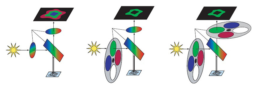
Why Traditional Light Sources Fall Short
Mercury and metal-halide lamps have long been used in fluorescence microscopy, but their limitations are well-documented:
Short Lifespan: Frequent bulb replacements (every 200–500 hours) increase costs and downtime.
Broad Emission Spectra: Unwanted wavelengths create background noise, forcing reliance on filter wheels that introduce mechanical vibrations.
Slow Shutter Speeds: Prolonged exposure accelerates photobleaching and phototoxicity, killing sensitive live cells.
Inflexibility: Fixed spectral lines limit compatibility with emerging fluorophores (e.g., far-red dyes).
These issues hinder advanced applications like super-resolution imaging, multiplexed assays, and longitudinal studies.
LED Light Sources: A Game-Changing Solution
Modern LED systems overcome traditional limitations while enhancing experimental control. Here’s how:
1. Precision Excitation with Narrow Spectra
LEDs emit light within 5–20 nm bandwidths, matching the absorption peaks of popular fluorophores (e.g., GFP, mCherry). This precision:
Reduces autofluorescence and crosstalk in multicolor experiments.
Eliminates filter wheels by pairing with multiband filters, minimizing vibrations.
2. Programmable Illumination for Complex Workflows
Millisecond switching: Rapidly toggle between wavelengths to image multiple labels without photodamage.
Adjustable intensity: Optimize excitation power for dim or photosensitive samples.
3. Enhanced Live-Cell Compatibility
Low heat output: LEDs generate minimal IR radiation, preserving cell viability during long-term imaging.
Reduced phototoxicity: Shorter exposure times (enabled by fast shutters) extend experiment durations.
4. Cost Efficiency and Sustainability
10,000+ hour lifespan: LEDs require no bulb replacements or recalibrations, cutting maintenance costs.
Energy-efficient: Consumes 70% less power than mercury lamps.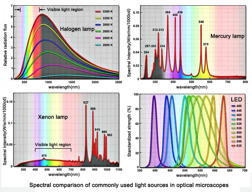
Custom LED Modules: Tailored for Your Research
As a leader in custom LED solutions, we design modules that align with your specific needs:
Multiplexed Imaging: Combine wavelengths (365nm, 488nm, 561nm, 640nm) for simultaneous 4-color detection.
Live-Cell Optimization: Integrate low-intensity modes and TTL triggering for gentle, synchronized imaging.
Third-Party Compatibility: Our SDK-supported modules work seamlessly with Nikon NIS, ZEN Blue, and MetaMorph® software.
Case Study: LEDs Transform Cardiac Cell Imaging
A 2023 study in Cell Reports demonstrated that switching to LED illumination increased SNR by 40% in beating cardiomyocyte imaging. Our clients report similar breakthroughs:
72-hour time-lapse imaging with 90% cell survival using low-phototoxicity protocols.
50% faster data acquisition in multiplexed tissue sections.
Upgrade to LED—Unlock Your Microscope’s Full Potential
Don’t let outdated light sources limit your science. Our custom LED modules empower you to:
Elimiate crosstalk with spectral precision.
Extend cell viability for critical live-cell studies.
Scale workflows with modular, software-controlled systems.
Contact us today to design a solution that elevates your imaging performance.


 Horticulture
 Uitraviolet
 Marine light